
Effective endocarditis symptoms, causes, complications
History
In patients with infective endocarditis (IE), the present illness history is highly variable. Symptoms commonly are vague, emphasizing constitutional complaints, or complaints may focus on primary cardiac effects or secondary embolic phenomena. Fever and chills are the most common symptoms; anorexia, weight loss, malaise, headache, myalgias, night sweats, shortness of breath, cough, or joint pains are common complaints as well.
Primary cardiac disease may present with signs of congestive heart failure due to valvular insufficiency. Secondary phenomena could include focal neurologic complaints due to an embolic stroke or back pain associated with vertebral osteomyelitis. As many as 20% of cases present with focal neurologic complaints and stroke syndromes.
Dyspnea, cough, and chest pain are common complaints of intravenous drug users. This is likely related to the predominance of tricuspid valve endocarditis in this group and secondary embolic showering of the pulmonary vasculature.
A key concern is the distinction between subacute and acute IE. The diagnosis of subacute IE is suggested by a history of an indolent process characterized by fever, fatigue, anorexia, back pain, and weight loss. Less common developments include a cerebrovascular accident or congestive heart failure.
The patient should be questioned about invasive procedures and recreational drug use that may be causing the bacteremia. Most subacute disease caused by S viridans infection is related to dental disease. Most cases are not caused by dental procedures but by transient bacteremias secondary to gingivitis. In 85% of patients, symptoms of endocarditis appear within 2 weeks of dental or other procedures.
The interval between the onset of disease and diagnosis averages approximately 6 weeks. The fact that less than 50% of patients have previously diagnosed underlying valvular disease significantly limits the effectiveness of antibiotic prophylaxis.
Acute IE is a much more aggressive disease. The patient notices an acute onset of high-grade fevers and chills and a rapid onset of congestive heart failure. Again, a history of antecedent procedures or illicit drug use must be investigated.
The distinction between these 2 polar types of IE has become less clear. Intermittent use of antibiotics aimed at treating misdiagnosed endocarditis can suppress bacterial growth within the valvular thrombus, giving rise to the state of muted IE. This is often the case in nosocomial infective endocarditis (NIE; also referred to as healthcare-associated IE [HCIE]), which commonly manifests with elements of a sepsis syndrome (ie, hypotension, metabolic acidosis fever, leukocytosis, and multiple organ failure).
The source of the bacteremia may be an infection in another organ (eg, pneumonia, pyelonephritis) or in a central venous catheter. Most often, these patients are in the intensive care unit. Approximately 45% of cases of NIE/HCIE occur in patients with prosthetic valves. Muted IE due to S aureus infection may resemble IE that results from S viridans infection.
Subacute native valve endocarditis
The symptoms of early subacute native valve endocarditis (NVE) are usually subtle and nonspecific. They include low-grade fever (absent in 3-15% of patients), anorexia, weight loss, influenzalike syndromes, polymyalgia-like syndromes, pleuritic pain, syndromes similar to rheumatic fever (eg, fever, dulled sensorium as in typhoid, headaches), and abdominal symptoms (eg, right upper quadrant pain, vomiting, postprandial distress, appendicitis-like symptoms).
When appropriate therapy is delayed for weeks or months, additional clinical features, embolic or immunological in origin, develop.
Signs and symptoms secondary to emboli include acute meningitis with sterile spinal fluid, hemiplegia in the distribution of the middle cerebral artery, regional infarcts that cause painless hematuria, infarction of the kidney or spleen, unilateral blindness caused by occlusion of a retinal artery, and myocardial infarction arising from embolization of a coronary artery.
The emboli of right-sided IE commonly produce pulmonary infarcts. The rate of embolization is related to the organism, the size of the vegetation and its rate of growth or resolution, and its location.
The vegetations of S aureus, Haemophilus influenzae, H parainfluenzae, and the fungi are much more likely to embolize than those of S viridans. Those larger than 10 mm in diameter and mobile or prolapsing have a high rate of embolization. A vegetation that grows during therapy is associated with a significant increase in the risk of embolization but with the persistence of bacteremia.
Clinically separating the importance of the absolute size and the rate of change in the size of the vegetation from the causative organism is difficult. The vegetations of the mitral valve are much more likely to embolize than those in any other location. The risk of embolization markedly decreases after 1 week of appropriate antibiotic therapy.
The deposition of circulating immune complexes in the kidney may produce interstitial nephritis or proliferative glomerulonephritis, with renal failure progressing to the point of uremia at the time of the patient’s presentation. Similarly, various musculoskeletal symptoms (44% of patients) arise from immunologically mediated synovitis.
Osler nodes and Roth spots arise from immune-mediated vasculitis. Patients may experience palpitations, ie, the symptoms of an immune-mediated myocarditis.
The origin of lumbosacral back pain in patients with subacute IE (15%) is unclear but probably results from the deposition of immune complexes in the disk space. However, antibiotic therapy rapidly abolishes these symptoms. In 50% of patients with cerebral emboli, this event is the first manifestation of IE and is associated with a 2- to 4-times higher mortality rate. Stroke in younger people should always raise the possibility of underlying IE.
Bacteria-free infective endocarditis
Rarely observed today, the bacteria-free state of IE is one in which patients have multiple negative blood culture results in the presence of severe congestive heart failure, renal failure, multiple sterile emboli, massive splenomegaly, severe anemia, brown facial pigmentation, bilateral thigh pain, and massive leg edema. These patients are usually afebrile. This process appears to indicate prolonged and unchecked stimulation of the immune system.
Acute infective endocarditis
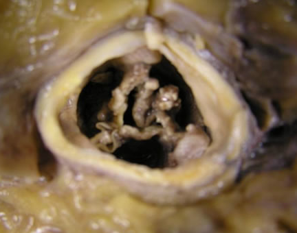
The clinical symptoms of acute IE result from either embolic or intracardiac suppurative complications. The onset of illness is abrupt, with rapidly progressive destruction of the infected valve (see the images below). The valvular leaflets are quickly destroyed by bacteria that multiply rapidly within the ever-growing friable vegetations. Complications develop within a week. These include the dyspnea and fatigue of severe congestive heart failure and a wide spectrum of neuropsychiatric complications resulting from CNS involvement.
 Acute bacterial endocarditis caused by Staphylococcus aureus with perforation of the aortic valve and aortic valve vegetations. Courtesy of Janet Jones, MD, Laboratory Service, Wichita Veterans Administration Medical Center.
 Acute bacterial endocarditis caused by Staphylococcus aureus with aortic valve ring abscess extending into myocardium. Courtesy of Janet Jones, MD, Laboratory Service, Wichita Veterans Administration Medical Center.
Intravenous-drug-abuse infective endocarditis
Patients with right-sided intravenous drug abuse (IVDA) IE (53% of cases) frequently present with pleuropulmonary (pneumonia and/or empyema) manifestations. Symptoms due to metastatic infection develop early in a disease course caused by S aureus. Right-sided disease is associated with a low rate of congestive heart failure and valvular perforation.
Infection with P aeruginosa has a high rate of neurological involvement, with 2 distinctive features: (1) mycotic aneurysms with a higher-than-average rate of rupture and (2) panophthalmitis (10% of patients). The course of infection with P aeruginosa is much slower than that of S aureus.
The course of left-sided IVDA IE is similar to that of non-IVDA disease.
Approximately 5-8% of febrile individuals who abuse intravenous drugs have underlying IE. Many users of illicit drugs may lose their fever within a few hours of hospitalization. This phenomenon, termed cotton wool fever, is probably caused by the presence of adulterants contained within the injected drugs.
Prosthetic valve endocarditis
Clinical features of prosthetic valve endocarditis (PVE) closely resemble those of NVE. Early PVE is defined as infection occurring within 60 days of valve implantation; late PVE occurs after this period. For valvular infection with coagulase-negative staphylococci (CoNS), this division should be extended to 12 months.
Congestive heart failure occurs earlier and is more severe in persons with PVE. The patient may present with symptoms of myocarditis or pericarditis. The rate of embolic stroke is high in the first 3 days of PVE.
Pacemaker infective endocarditis
The clinical presentation in a person with a pacemaker infection and pacemaker IE depends on several factors, including the site of infection (eg, generator pocket vs intravascular leads or epicardial leads), the type of organism, and the origin of the infection (eg, pocket erosion, localized infection of the generator pocket, bacteremia from a remote site).
Early infections, within a few months of implantation, manifest as acute or subacute infections of the pulse-generator pocket. Bacteremia may be present even in the absence of clinical signs and symptoms. Fever is the most common finding and may be the only finding in approximately 33% of patients.
Late infections of the pocket may be due to erosion of the overlying skin without systemic involvement. Such erosions always indicate infection of the underlying device.
The most significant late infections involve the transvenous or epicardial leads. With epicardial infection, signs and symptoms of pericarditis or mediastinitis may be present along with bacteremia. Infection of the transvenous electrode produces signs and symptoms of right-sided endocarditis. Those that occur early after implantation (33% of cases) show prominent systemic signs of infection, often with obvious localization to the pacemaker pocket.
Late infections have much more subtle manifestations. They may occur up to several years after implantation or reimplantation.
Fever is almost universal in persons with pacemaker IE. Signs of right-sided endocarditis (ie, pneumonia, septic emboli) are observed in up to 50% of patients.
Nosocomial infective endocarditis
NIE commonly manifests with elements of a sepsis syndrome (ie, hypotension, metabolic acidosis fever, leukocytosis, and multiple organ failure). The source of bacteremia may develop from an infection in another organ (eg, pneumonia, pyelonephritis) or from a central venous catheter. Most often, these patients are in the intensive care unit.
Approximately 45% of cases of NIE/HCIE occur in patients with prosthetic valves.
Physical Examination
General findings
Fever, possibly low-grade and intermittent, is present in 90% of patients.
The AHA (endorsed by the Infectious Diseases Society of America [IDSA]) 2010 guideline update on cardiovascular implantable electronic device (CIED) infections and their management recommends that patients with CIED who develop unexplained fever or bloodstream infection should seek evaluation for CIED infection by cardiologists or infectious disease specialists. [35]
Heart murmurs are heard in approximately 85% of patients. Change in the characteristics of a previously noted murmur occurs in 10% of these patients and increases the likelihood of secondary congestive heart failure.
One or more classic signs of IE are found in as many as 50% of patients. They include the following:
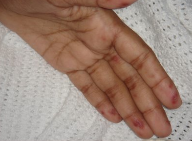
- Petechiae - Common but nonspecific finding (see the image below)
- Subungual (splinter) hemorrhages - Dark red linear lesions in the nailbeds
- Osler nodes - Tender subcutaneous nodules usually found on the distal pads of the digits
- Janeway lesions - Nontender maculae on the palms and soles
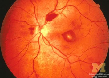
- Roth spots - Retinal hemorrhages with small, clear centers; rare and observed in only 5% of patients.
A middle-aged man with a history of intravenous drug use who presented with severe myalgias and a petechial rash. He was diagnosed with right-sided staphylococcal endocarditis.
Signs of neurologic disease occur in as many as 40% of patients. Embolic stroke with focal neurologic deficits is the most common etiology. Other etiologies include intracerebral hemorrhage and multiple microabscesses. [1]
Signs of systemic septic emboli are due to left heart disease and are more commonly associated with mitral valve vegetations. Multiple embolic pulmonary infections or infarctions are due to right heart disease.
Signs of congestive heart failure, such as distended neck veins, frequently are due to acute left-sided valvular insufficiency.
Splenomegaly may be present.
Other signs include the following:
- Stiff neck
- Delirium
- Paralysis, hemiparesis, aphasia
- Conjunctival hemorrhage
- Pallor
- Gallops
- Rales
- Cardiac arrhythmia
- Pericardial rub
- Pleural friction rub
Subacute infective endocarditis
Approximately 3-15% of patients with subacute IE (primarily elderly and chronically ill individuals) have normal or subnormal temperatures. The vast majority of patients have detectable heart murmurs. The presence of a murmur is so common (99% of cases) that its absence should cause clinicians to reconsider the diagnosis of IE. The major exception is right-sided IE, in which only one third of patients have a detectable murmur.
Because many of these murmurs are hemodynamically insignificant and have been present for years, their role in the patient’s illness may be underestimated. The saying "a changing murmur is extremely helpful in diagnosing subacute IE" is a myth. Only 15% do so early in the course of infection.
The peripheral lesions of subacute IE are observed in only approximately 20% of patients, compared with 85% in the preantibiotic era. Currently, the most common of these is petechiae. They may occur on the palpebral conjunctivae, the dorsa of the hands and feet, the anterior chest and abdominal walls, the oral mucosa, and the soft palate.
Subungual hemorrhages (ie, splinter hemorrhages) are linear and red. They are usually caused by workplace trauma to the hands and feet rather than by valvular infection. Hemorrhages that do not extend for the entire length of the nail are more likely the result of infection rather than trauma.
Osler nodes are smallish tender nodules that range from red to purple and are located primarily in the pulp spaces of the terminal phalanges of the fingers and toes, soles of the feet, and the thenar and hypothenar eminences of the hands. Their appearance is often preceded by neuropathic pain. They last from hours to several days. They remain tender for a maximum of 2 days. The underlying mechanism is probably the circulating immunocomplexes of subacute IE. They have been described in various noninfectious vasculitides.
Clubbing of fingers and toes was found almost universally, but it is now observed in less than 10% of patients. It primarily occurs in those patients who have an extended course of untreated IE.
The arthritis associated with subacute IE is asymmetrical and is limited to 1-3 joints. Clinically, it resembles the joint changes found in patients with rheumatoid arthritis, Reiter syndrome, or Lyme disease. The fluid is usually sterile.
Splenomegaly is observed more commonly in patients with long-standing subacute disease. It may persist long after successful therapy.
Roth spots are retinal hemorrhages with pale centers. The Litten sign represents cotton-wool exudates.
Acute infective endocarditis
In approximately one third of patients with acute IE, murmurs are absent. The most common type is an aortic regurgitation murmur. Because of the suddenness of onset, the left ventricle does not have a chance to dilate. In this situation, the classic finding of increased pulse pressure in significant valvular insufficiency is absent.
Fever is always present, and it usually is high.
Janeway lesions are irregular erythematosus and painless macules (1-4 mm in diameter). They most often are located on the thenar and hypothenar eminences of the hands and feet. They usually represent an infectious vasculitis of acute IE resulting from S aureus infection.
Acute septic monoarticular arthritis in patients with acute IE most often is caused by S aureus infection.
Complications
The following are potential complications of IE:
- Myocardial infarction, pericarditis, cardiac arrhythmia
- Cardiac valvular insufficiency
- Congestive heart failure
- Sinus of Valsalva aneurysm
- Aortic root or myocardial abscesses
- Arterial emboli, infarcts, mycotic aneurysms
Arthritis, myositis- Glomerulonephritis, acute renal failure
- Stroke syndromes
- Mesenteric or splenic abscess or infarct[52]
Congestive heart failure due to aortic valve insufficiency is the most common intracardiac complication of subacute endocarditis. It develops after months of untreated disease but may occur a full year following microbiological cure.
The complication of arterial embolization is second in frequency to congestive heart failure for both subacute and acute IE. The frequency of this complication has decreased, from 80% in the preantibiotic era to 15-35% today. The emboli are usually sterile because of the minimally invasive nature of the causative organisms (eg, S viridans).
The persons most at risk are younger (20-40 y), have mitral or aortic valve (native or prosthetic) involvement, and are infected with certain organisms such as Candida or Aspergillus species, S aureus, Haemophilus parainfluenzae, group B streptococci, and nutritionally variant streptococci.
The prevalence of embolization appears to be the same for both types of disease. The most common areas of deposition include the coronary arteries, kidneys, brain, and spleen. Infarction at the site of embolization is common; abscess formation is not. Cerebral emboli occur in 33% of patients. The middle cerebral artery is involved most often.
Other neurological embolic damage includes cranial nerve palsies, cerebritis, and mycotic aneurysms caused by weakening of the vessel walls and produced by embolization to the vasa vasorum. Mycotic aneurysms may occur in the abdominal aorta and the splenic, coronary, and pulmonary arteries.
In acute IE, the frequency of aneurysms and other suppurative intracardiac complications is high. In addition to valvular insufficiency, other intracardiac complications of acute IE include (1) aortocardiac and other fistulas, (2) aneurysms of the sinus of Valsalva, (3) intraventricular abscesses, (4) ring abscesses, (5) myocardial abscesses, (6) mycotic aneurysms, (7) septic coronary arterial emboli, and (8) pericarditis.
In patients with acute disease, especially disease caused by S aureus infection, emboli almost inevitably lead to abscesses in the areas where they are deposited. Multiple abscesses can occur in almost every organ, including the kidneys, heart, and brain. Mycotic aneurysms may occur in almost any artery. Paradoxically, they are less common in patients with acute IE. [53, 54]
It appears that older patients have higher rates of myocardial infarction and death but lower rates of neurological complications. [24]
Diagnostic Considerations
Definitive diagnosis of infective endocarditis (IE) is generally made by using the Duke criteria.
Duke diagnostic criteria
Durack and colleagues developed diagnostic criteria that combine the clinical, microbiological, pathological, and echocardiographic characteristics of a specific case. [2]
Major blood culture criteria include the following:
- Two blood cultures positive for organisms typically found in patients with IE (ie, S viridans, Streptococcus bovis, a HACEK group organism, community-acquired S aureus, or enterococci in the absence of a primary focus)
- Blood cultures persistently positive for one of the above organisms from cultures drawn more than 12 hours apart
- Three or more separate blood cultures drawn at least 1 hour apart
Major echocardiographic criteria include the following:
- Echocardiogram positive for IE, documented by an oscillating intracardiac mass on a valve or on supporting structures, in the path of regurgitant jets, or on implanted material in the absence of an alternative anatomical explanation
- Myocardial abscess
- Development of partial dehiscence of a prosthetic valve
- New-onset valvular regurgitation
Minor criteria include the following:
- Predisposing heart condition or intravenous drug use
- Fever of 38°C (100.4°F) or higher
- Vascular phenomenon, including major arterial emboli, septic pulmonary infarcts, mycotic aneurysm, intracranial hemorrhage, conjunctival hemorrhage, or Janeway lesions
- Immunological phenomenon such as glomerulonephritis, Osler nodes, Roth spots, and rheumatoid factor
- Positive blood culture results not meeting major criteria or serologic evidence of active infection with an organism consistent with IE (eg, Brucella, C burnetii [ie, Q fever], Legionella)
- Echocardiogram results consistent with IE but not meeting major echocardiographic criteria
Definitive pathological diagnosis is established by demonstrating microorganisms, by culture or histology, in vegetations removed by surgery, embolectomy, or drainage of an intracardiac abscess. Alternatively, a definitive clinical diagnosis is made based on the presence of 2 major criteria, 1 major criterion and 3 minor criteria, or by 5 minor criteria.
A diagnosis of possible IE is made when findings consistent with IE fall short of the criteria for definite IE but do not meet the criteria for rejection.
Rejection criteria for the diagnosis of IE are as follows:
- The presence of a firm alternative diagnosis of the manifestations of endocarditis
- Resolution of manifestations of endocarditis after 4 or fewer days of antimicrobial therapy
- No pathologic evidence of IE at surgery or autopsy after 4 or fewer days of antimicrobial therapy
These criteria may, at times, overdiagnose IE and may not be as applicable in patients with subacute disease.
Other problems to be considered include the following:
- Thrombotic nonbacterial endocarditis
- Vasculitis
- Temporal arteritis
- Marantic endocarditis
- Connective tissue disease
- Fever of unknown origin (FUO)
- Intra-abdominal infections
- Septic pulmonary infarction
- Tricuspid regurgitation


Post a Comment